Research Summary: Assessing Organisms in Great Lakes Ballast Water
0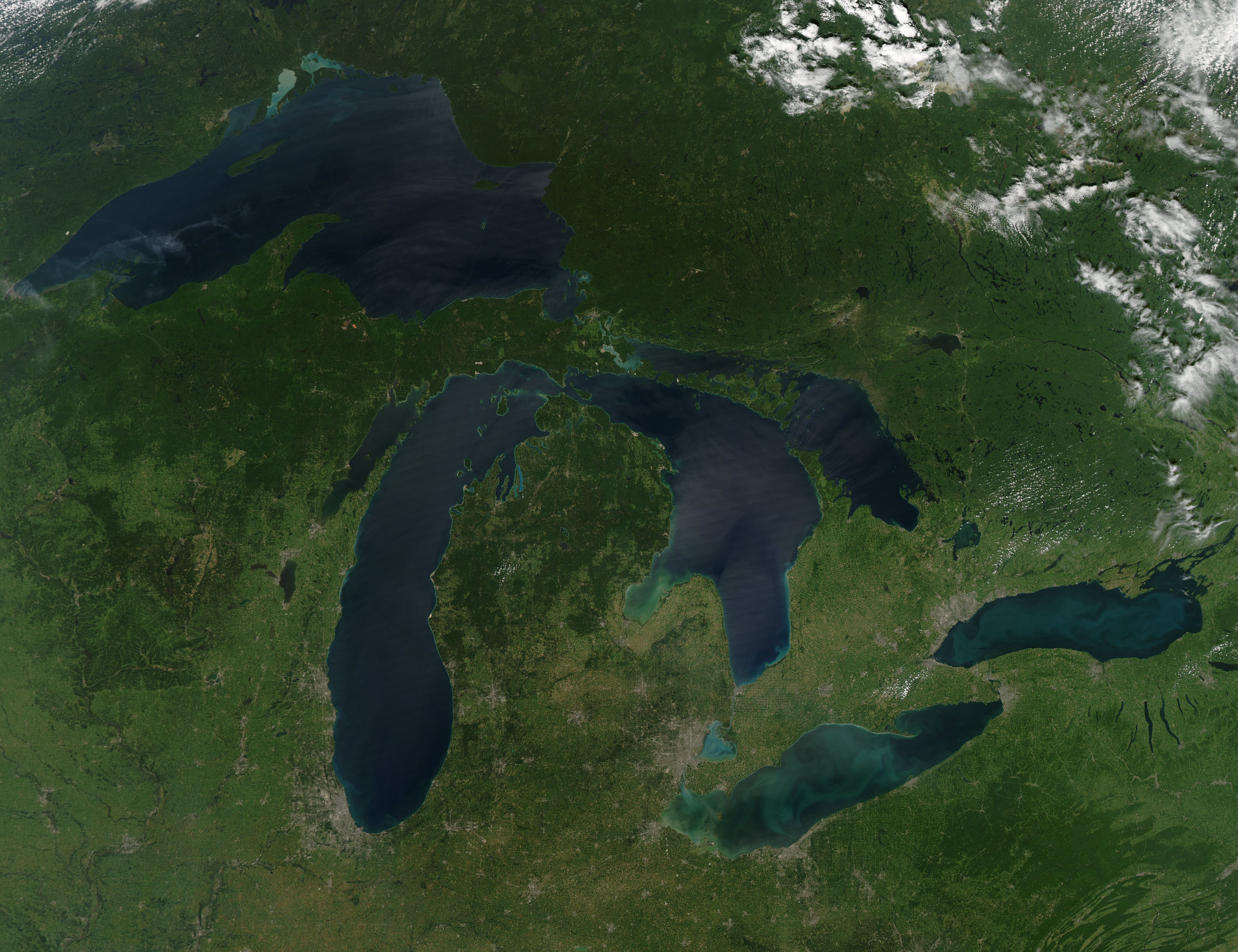
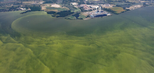
Great Lakes region on a cloudless summer day, August 2010. (Credit: NASA Earth Observatory)
For decades the Great Lakes have been subject to invasive species introductions through the discharge of ships’ ballast water. Several treatment technologies involving physical, chemical, and biological processes have been developed to remove or inactivate organisms in this discharge. Assessing the efficacy of these technologies involves estimating the number of viable propagules in treated discharge relative to untreated controls. For organisms in the 10–50 μm (microns) size range, for example, the International Maritime Organization (IMO) mandates that fewer than 10 viable organisms per milliliter may be discharged. To date, however, there is no standard method to assess viability of natural assemblages of organisms in this size group (largely phytoplankton and protozoans) in freshwater environments. We report here on a process of assemblage concentration, staining with fluorescein diacetate (FDA), and microscopic observation as a reliable and efficient method to assess densities of viable freshwater organisms in this size category in ballast discharge. A number of other methods, including digestion with enzymes, flow cytometry, and a variety of vital and mortal stains, were tested and discarded during this vetting process due to inconsistent or ambiguous results.
Methods
Using available literature, possible methods for quantitatively discerning living and dead organisms less than 50 μm and greater than or equal to 10 μm in minimum dimension were reviewed against a set of criteria for their potential applicability to mixed unknown assemblages associated with freshwater ballast discharge. The criteria for evaluation of each method were:
1. Signal is quantitatively precise such as that necessary to assess organism condition upon discharge from a ship’s ballast system (lack of false negatives or positives at the time of analysis);
2. Signal is consistent across taxa in ambient freshwater assemblages;
3. Signal is consistent across treatment types;
4. Method is practical for rapid, high throughput, on-site assessments.
Methods considered were (1) natural autofluorescence from chlorophyll to identify living cells; (2) vital, metabolic, and mortal stains such as those typically associated with specific standard test organisms including propidium iodide, fluorescein diacetate, Sytox® Green and several stain combinations; (3) cell digestion methods, particularly DNAse + trypsin digestion; and (4) automated cell enumeration methods including standard flow cytometry combined with use of vital and mortal stains.

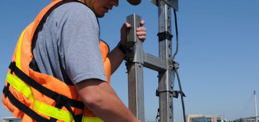
Apparatus used for concentration of organism assemblages in whole-water samples. (Credit: Euan Reavie)
The subset of methods that, based on available literature, were determined to potentially meet criteria outlined above was empirically validated against the same criteria using ambient freshwater assemblages of organisms. Six viability analysis methods (five epifluorescent staining methods and one cell digestion method) and one flow cytometry method for automated counting and characterization of prepared samples were investigated. All quantitative measures were maintained throughout evaluations, including volumes filtered, backwashed and strewn on counting chambers, total lengths and widths of microscopic transects, and cell and entity counts.
Results
This study reviewed and tested several methods in order to choose the best one for determining viability of freshwater microorganism assemblages occurring in treated ballast water. From these analyses, microscopic examination of FDA-stained samples appears to be the best approach currently available for determining viability, while
fluorescence microscopy remains the best method for live cell enumeration. Autofluorescence gives frequent false positives and therefore has been discounted as a viability determination method in previous studies.
Based on our empirical investigations of candidate stains, we recommend caution when using Sytox and the LIVE/DEAD kits to perform viability assessments in natural freshwater assemblages. Our research indicated clear discrepancies between the reported ability to discern living from dead organisms and the actual method performance in laboratory tests. We believe this discrepancy arose because methods were originally designed for application to (and tested with) standard laboratory test organisms, and not natural assemblages. Also of note is the fact that stains rejected by this study were not adequately quantitative for this application, in general. While most staining methods can categorically distinguish living from dead cells, in an application in which the precise number of live is sought in a regulatory context, the proportion of incorrect or ambiguous results precludes their use. This observation, however, does not preclude their use for other types of ballast treatment research. For example, Sytox Green accurately measures mortality in some monocultures, and we found it to be a highly reliable method for bench-scale assessments using cultured Selenastrum capricornutum following treatment applications (unpublished data). We rejected the cell digestion method due to our inability to reliably digest dead cells. However, the problems we confronted might be specific to northern Minnesota and Lake Superior water conditions, rather than freshwater generally. The natural assemblages and water matrix tested in this study may be less susceptible to artificial lysis with DNAse and trypsin. This method has potential to provide rapid cost-effective viability assessments and should be studied and developed for possible use across a wider variety of environments and assemblages.
We found that assemblage concentration, staining with FDA, and microscopic observation met our evaluation criteria as a reliable method to discern live densities of viable freshwater organisms in the 10–50 μm size category in ballast discharge. Though FDA may be inappropriate for some environments and species assemblages (one researcher suggests that it does not adequately stain some diatoms from coastal marine environments; Mario Tamburri, Maritime Environmental Resource Center, Chesapeake Biological Laboratory, personal communication), we are confident that it is suitable to ballast water treatment assessment in Lake Superior. An initial phase
of method evaluation is recommended for any testing program to validate its use in a particular subsystem within the Great Lakes. It should be noted that any staining method, even one that appears to have broad spectrum applicability and reliability across treatment processes (like FDA did in this study) may not discern living from viable cells. There are probably many cases where ballast water treatment systems render living cells no longer reproductively viable, and thereby eliminate any environmental risk associated with those cells. However, confirmation that living cells are viable would require subsequent grow-out experiments following treatment.

Array of results for various treatments and viability assessment methods for natural assemblages in the 10–50 μm size group. Proportions of live, dead, and ambiguous cells for each treatment are based on the average of at least five sample assessments. Significance of comparisons are based on at least five assemblage counts for each treatment using a nonparametric Student’s t-test between numbers of (apparently) living cells in treated and untreated (control) assemblages. (Credit: Euan Reavie)
Grow-out is especially important if testing includes resting cysts, such as those from amoebae, chrysophytes, and dinoflagellates. To make conservative risk assessments, regulators are likely to assume that cells deemed to be living via a reliable stain are also able to propagate if introduced to an aquatic environment. With regard to characterizing numbers of live cells, we see no reliable substitute for microscopy. In particular, the ability of flow cytometry to automate counting and sizing of live cells in ambient assemblages is limited by the stain reliability as noted above.
Accordingly, flow cytometry, including FlowCAM, appears ideally suited to assessing viability in monocultures and unicellular entities (Cid et al., 1996). Microscopy, however, may limit sample volume necessary to achieve statistical confidence if regulatory criteria designate analysis of larger sample volumes. Realistically, an approximate limit for one analyst using the microscopic method would be complete analysis of 3 ml of sample water at 1000 living cells/ml within 1 h, subject to the complexity of the species assemblage.
The selected method appears capable of performing the task of live/dead protist enumeration in natural freshwater assemblages. However, ship discharge monitoring imposes additional requirements on analytical tools. Variable ballast water sources such as occur in ships greatly complicate the tasks, and there may be unanticipated
influences by certain treatment methods on the effectiveness of FDA in vitality assessments. Nonetheless, we have eliminated several methods that are currently not suited to ballast assessments in the Great Lakes, and the selected method provides a strong foundation for future microorganism assessments in ballast water.
Acknowledgements
We thank Fluid Imaging Technologies for support during the flow cytometry workshop. Kathleen Kennedy helped with field collections. This work was supported by funds and in-kind efforts assembled from the private sector, federal grants, Congressional appropriations, foundations, and states, including contributions from Canadian and
US Great Lakes ports, National Oceanic and Atmospheric Administration, St. Lawrence Seaway Development Corporation, St. Lawrence Seaway Management Corporation, US Maritime Administration, US Department of Transportation, Legislative-Citizen Commission on Minnesota Resources, University of Wisconsin Superior (Balcer M,
TenEyck M), University of Minnesota Duluth (Hicks R, Branstrator D), Great Lakes carrier companies, and the Great Lakes Maritime Research Institute. This is contribution number 501 of the Center for Water and the Environment, Natural Resources Research Institute, University of Minnesota Duluth.
Full study and references published September 2010 in the Journal of Great Lakes Research (Volume 36, Issue 3, pages 540-547).
Selected References
- Agustí, S., 2004. Viability and niche segregation of Prochlorococcus and Synechcoccus cells across the central Atlantic ocean. Aquat. Microb. Ecol. 36, 53–59.
- Agustí, S., Sánchez, C.M., 2002. Cell viability in natural phytoplankton communities quantified by a membrane permeability probe. Limnol. Oceanogr. 47, 818–828.
- Agustí, S., Alou, E., Hoyer, M.V., Frazer, T.K., Canfield, D.E., 2006. Cell death in lake phytoplankton communities. Freshw. Biol. 51, 1496–1506.
- Berglund, D.L., Taffs, R.E., Robertson, N.P., 1987. A rapid analytical technique for flow cytometric analysis of cell viability using calcofluor white m2r. Cytometry 8,
421–426. - Booth, B.C., 1987. The use of autofluorescence for analyzing oceanic phytoplankton communities. Bot. Marina 30, 101–108.
- Bricelj, V.M., Lonsdale, D.J., 1997. Aureococcus anophagefferens: causes and ecological consequences of brown tides in U.S. Mid-Atlantic coastal waters. Limnol. Oceanogr.
42, 1023–1038. - Buggé, D.M., Allam, B., 2005. A fluorometric technique for the in vitro measurement of growth and viability of quahog parasite unknown (qpx). J. Shellfish Res. 24,
1013–1018. - Buskey, E.J., Hyatt, C.J., 2006. Use of the FlowCAM for semi-automated recognition and enumeration of red tide cells (Karenia brevis) in natural plankton samples. Harmful Algae 5, 685–692.
- Buttino, I., do Santo Espirito, M., Ianora, A., Miralto, A., 2004. Rapid assessment of copepod (Calanus helgolandicus) embryo viability using fluorescent probes. Mar.
Biol. 145, 393–399. - Cangelosi, A., Mays, N., 2005. Summary and Findings of the Ballast Discharge Monitoring Device Workshop, 2002 Aug. 12–16; U.S. Geological Survey’s Western
Fisheries Research Center’s Marrowstone Island Field Station, WA. Northeast- Midwest Institute, Washington, DC. 53 pp. (http://www.nemw.org/images/stories/documents/marrowstonereport.pdf).


